Chest CT scans are widely used for many medical reasons, including lung-cancer screening.
But the esophagus has traditionally been difficult to assess for small early cancers on ordinary non-contrast CT images.
A newly published artificial-intelligence system may help change that.
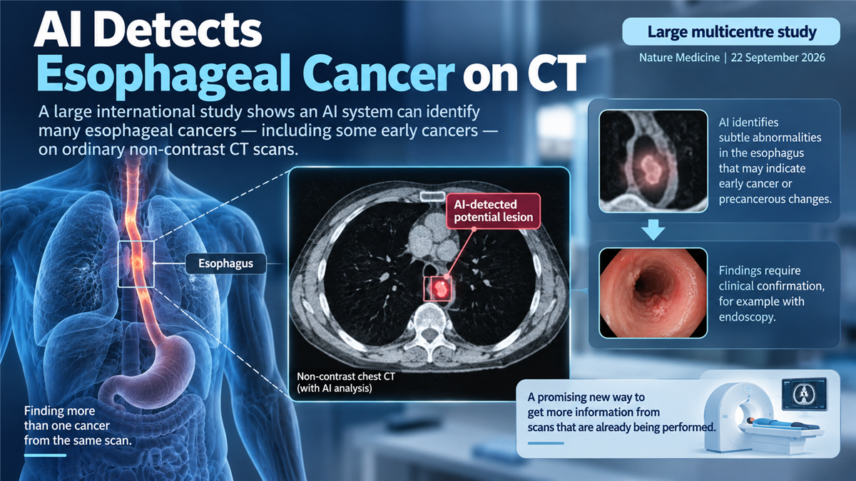
Researchers have developed an AI system called EAGLE — Esophageal AI-Guided malignant Lesion Evaluation — designed to detect esophageal cancer and precancerous lesions using non-contrast CT scans.
In a large multicentre study published in Nature Medicine on 22 September 2026, the system was evaluated across diverse clinical settings and populations.
The overall research involved more than 80,000 people.
The findings suggest that AI may be able to extract useful esophageal-cancer information from CT scans that were not traditionally considered reliable screening tools for small esophageal lesions.
However, the technology is not a replacement for endoscopy, and the study does not establish that AI-based CT screening reduces deaths from esophageal cancer.
Why is esophageal cancer difficult to detect early?
Esophageal cancer can be difficult to identify before symptoms develop.
Early lesions may be small and cause few or no symptoms.
Endoscopy allows doctors to directly inspect the lining of the esophagus and obtain tissue samples when necessary.
It is therefore an important diagnostic tool.
But endoscopy is invasive, requires specialist resources and is not practical as a universal screening procedure in many populations.
CT scanning is far more widely performed.
The problem is that the esophagus is a hollow structure that can collapse and move, producing imaging appearances that make small lesions difficult to distinguish from normal tissue.
Non-contrast CT is particularly challenging for this purpose.
What is EAGLE?
EAGLE is an artificial-intelligence system trained to analyse non-contrast CT images for patterns associated with malignant or precancerous esophageal lesions.
The model was developed using data from 6,813 patients.
Researchers then evaluated it in multiple independent settings rather than relying only on the data used to develop the model.
Across the overall research programme, data came from 12 centres in three countries and involved approximately 80,612 people.
That scale and the use of external, real-world and prospective cohorts strengthen the evidence compared with an AI model tested only retrospectively at the hospital where it was developed.
How well did it perform?
In multicentre external testing involving 11,466 people, the system achieved approximately:
- 98.5% specificity
- 90.0% sensitivity for esophageal cancer
- and 52.5% sensitivity for precancerous lesions.
These figures need careful interpretation.
The high specificity means the system correctly classified most people without the target disease.
The 90% cancer sensitivity is encouraging.
But the considerably lower sensitivity for precancerous lesions means many such lesions were still missed.
Was the system tested in real clinical settings?
Yes.
The research included several additional validation settings.
A real-world calibration cohort included more than 35,000 people.
Prospective hospital validation involved more than 17,000 people.
A further real-world cohort involving low-dose CT screening included more than 10,000 people.
Researchers also examined cohorts in which CT findings could be compared with endoscopy.
At a threshold selected to favour greater sensitivity, the system detected approximately:
- 65.0% of precancerous lesions
- and 78.4% of stage I cancers
in the relevant paired CT/endoscopy cohorts.
This suggests the technology may identify lesions that conventional interpretation of non-contrast CT could overlook.
Why could this be useful?
One attractive possibility is that the technology could obtain additional information from CT scans that are already being performed.
For example, low-dose chest CT is used in some populations for lung-cancer screening.
If an AI system could also reliably flag people who may have an esophageal lesion requiring further investigation, the same scan could potentially provide information about more than one disease.
That differs from introducing an entirely new screening procedure.
It may therefore offer a scalable way to identify people who should undergo more definitive testing.
Does AI diagnose the cancer?
No.
An AI flag on a CT scan should not be considered a definitive cancer diagnosis.
Suspicious findings require appropriate clinical assessment and, when indicated, endoscopy and biopsy.
A diagnostic system can help identify people who may need further investigation.
That is different from independently establishing a cancer diagnosis.
Can CT now replace endoscopy?
No.
The study does not establish non-contrast CT as a replacement for endoscopy.
Endoscopy provides direct visualization of the esophageal lining and allows tissue sampling.
The AI system's sensitivity also varied according to lesion type, disease stage and operating threshold.
Precancerous lesions were particularly challenging.
A system that misses a substantial proportion of precancerous disease cannot simply replace a more definitive diagnostic method.
Does this prove that AI screening saves lives?
No.
This study primarily evaluates diagnostic performance.
Showing that an AI system detects cancers accurately is not the same as showing that using it as a screening programme reduces deaths.
To establish that, researchers would need to determine whether introducing the system into clinical practice:
- leads to earlier meaningful diagnosis
- improves treatment outcomes
- reduces esophageal-cancer mortality
- avoids excessive unnecessary endoscopies
- and remains accurate across different populations and healthcare systems.
Those questions require further prospective clinical evaluation.
Why is the study important?
Many medical AI studies perform well on carefully selected retrospective datasets but struggle when moved into different hospitals or real clinical practice.
The EAGLE study is notable because the system was evaluated across multiple centres, countries and large real-world and prospective cohorts.
It also tackles a clinically difficult problem: identifying subtle esophageal lesions on a type of scan that is already widely available.
The concept is therefore not simply "AI reads a scan."
It is that AI might allow clinicians to obtain new diagnostic information from existing imaging.
That could become particularly valuable if future studies demonstrate that the approach improves clinically important outcomes.
What happens next?
Further studies will need to determine:
- how the system performs in additional countries and populations
- how it affects endoscopy referrals
- the balance between missed lesions and unnecessary investigations
- how radiologists should integrate AI alerts into routine practice
- whether it improves early diagnosis
- and ultimately whether its use reduces deaths from esophageal cancer.
For now, the evidence supports a promising but appropriately limited conclusion:
AI can identify many esophageal cancers on ordinary non-contrast CT scans with high specificity across large and diverse study populations, but it remains a detection tool requiring clinical confirmation rather than a replacement for endoscopy or a proven mortality-reducing screening programme.